Internal View Of Heart | A healthy heart supplies your body with the right amount of blood at the rate needed to work well. The internal wall of the right atrium is composed of a smooth posterior portion (into which the vena cavae and coronary sinus drain) and a ridgelike, muscular anterior portion. Don't forget to share this picture with others via facebook, twitter, pinterest or other social medias! Lobes of the brain (superior view). Your heart's electrical system controls the rate and rhythm of your heartbeat.
Your heart's electrical system controls the rate and rhythm of your heartbeat. Internal features rt atrium consists of 1. The heart is a muscular organ about the size of a closed fist that functions as the body's circulatory pump. It takes in deoxygenated blood through the veins and delivers it to the lungs for oxygenation before pumping it into the various arteries (which provide oxygen and nutrients to body tissues by. Image depicting several anatomical landmarks of internal cardiac anatomy.
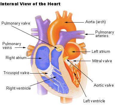
The pumped blood carries oxygen and nutrients to the body, while carrying metabolic waste such as carbon dioxide to the lungs. During the normal cardiac cycle, the right atrium receives deoxygenated blood from the body.once both atria are full, they contract, and the deoxygenated blood from the right atrium flows into the. Find the perfect internal view heart stock photo. Intraoperatively, the anatomy of the heart is viewed from the right side of the supine patient via a median sternotomy incision. Describe the internal features of right atrium. Heart presentation by slapadula 103512 views. List the chambers of the heart. The heart is a muscular organ about the size of a closed fist that functions as the body's circulatory pump. Your heart's electrical system controls the rate and rhythm of your heartbeat. It is the right upper chamber of the heart and receives venous blood from whole body pumps it to the right ventricle through right atrio ventricular or tricuspid. A wire electrode is attached to the fetal scalp or internal uterine pressure monitoring is sometimes used along with internal fetal heart rate monitoring. The external surfaces of the valves are covered by endocardium. Free online quiz internal view of the heart.
The heart is one of the most (internal structure of a heart) important organ of human body. The walls of the ventricles are relatively thicker than atrial walls. List the chambers of the heart. The heart is divided into a right and left side by the septum. Start studying internal view of heart.
![]()
Free online quiz internal view of the heart. The term cardiac (as in cardiology) means related to the heart and comes from the. Internal view of the human heart. The heart has four chambers, two relatively small upper chambers called atria and two larger lower chambers called ventricles. On its surface, it has the heart has been described by many texts as a pyramid which has fallen over. The upper right chamber of the heart. Recall that the heart's contraction cycle follows a dual pattern of circulation—the pulmonary (lungs)and systemic (body) circuits—because of the pairs of in order to develop a more precise understanding of cardiac function, it is first necessary to explore the internal anatomical structures in more detail. The heart is a muscular organ about the size of a closed fist that functions as the body's circulatory pump. Describe the internal features of right atrium. It takes in deoxygenated blood through the veins and delivers it to the lungs for oxygenation before pumping it into the various arteries (which provide oxygen and nutrients to body tissues by. Lobes of the brain (superior view). Thank you for visiting images of internal structure of heart pictures. A wire electrode is attached to the fetal scalp or internal uterine pressure monitoring is sometimes used along with internal fetal heart rate monitoring.
Free online quiz internal view of the heart. I think one of the best ways to understand the internal structures of the heart is by learning the passage of blood flow through the heart! Anterior view of the heart. Different structures of heart right atrium: This hd wallpaper images of internal structure of heart has viewed by 794 users.
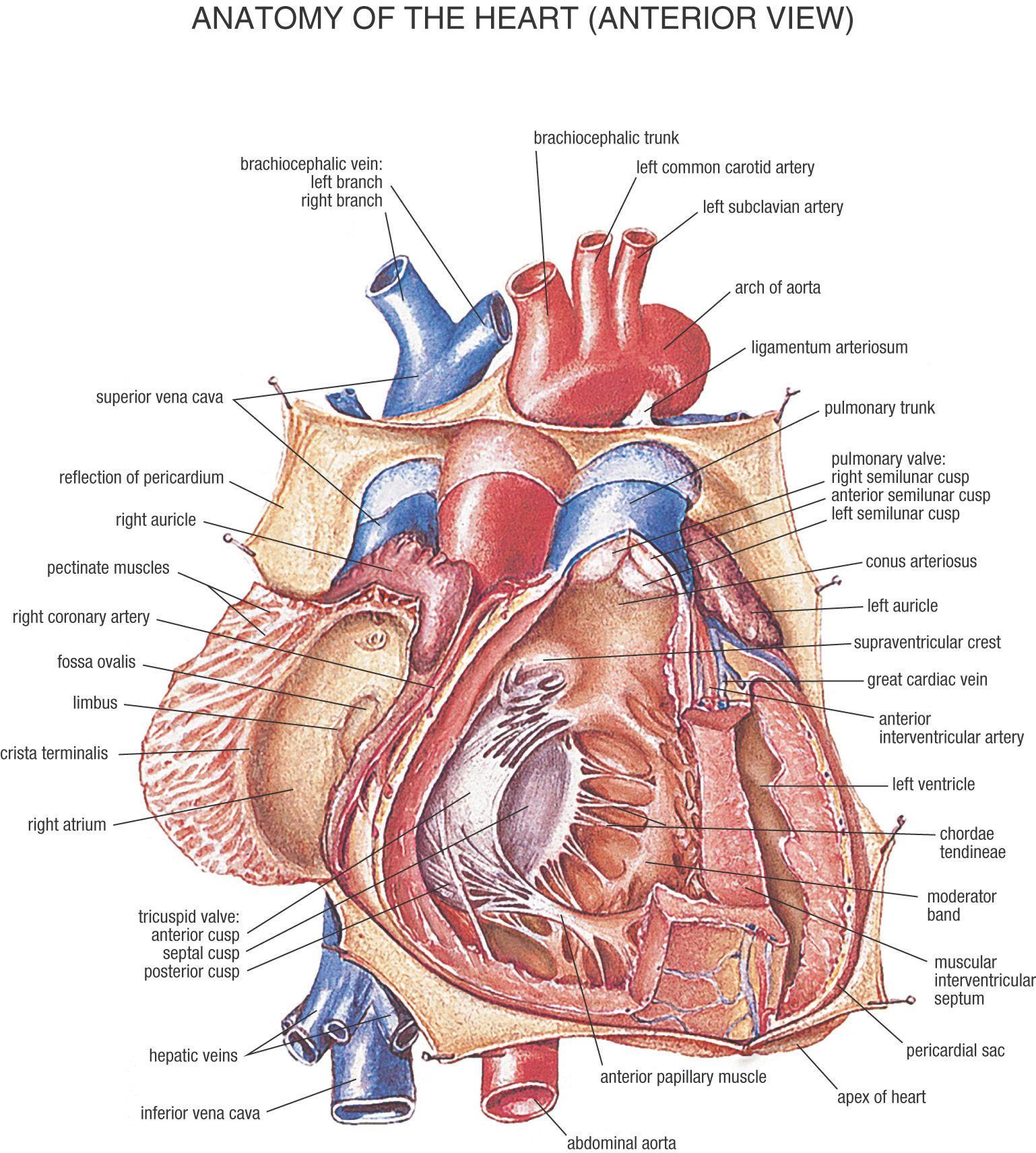
Intraoperatively, the anatomy of the heart is viewed from the right side of the supine patient via a median sternotomy incision. This view labelled illustration is from 'asklepios atlas of the human anatomy'. To establish the etiology of heart failure in patients with congenital heart disease can be challenging. Lobes of the brain (superior view). They are composed mostly of fibrous connective tissue that extends from the heart walls. During the normal cardiac cycle, the right atrium receives deoxygenated blood from the body.once both atria are full, they contract, and the deoxygenated blood from the right atrium flows into the. Start studying internal view of heart. On its surface, it has the heart has been described by many texts as a pyramid which has fallen over. Thank you for visiting images of internal structure of heart pictures. The internal wall of the right atrium is composed of a smooth posterior portion (into which the vena cavae and coronary sinus drain) and a ridgelike, muscular anterior portion. Anterior view of the heart. Your heart's electrical system controls the rate and rhythm of your heartbeat. Image depicting several anatomical landmarks of internal cardiac anatomy.
Internal View Of Heart: A wire electrode is attached to the fetal scalp or internal uterine pressure monitoring is sometimes used along with internal fetal heart rate monitoring.
Source: Internal View Of Heart
0 comments:
Post a Comment